
FIGURE 2.
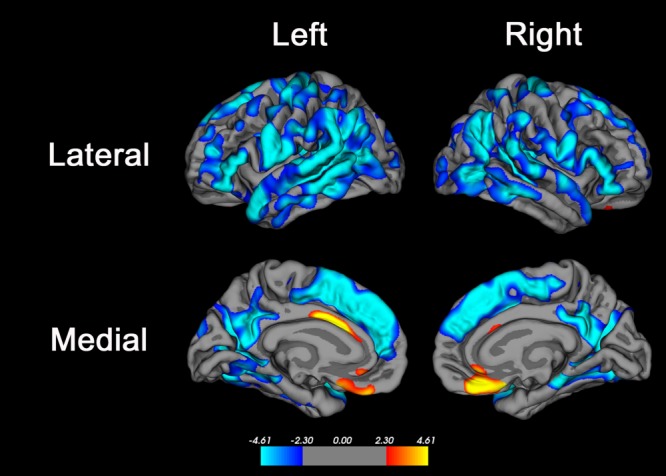
Brain regions where age was associated with cortical thickness. Advanced age was associated with decreased cortical thickness in many brain regions, most significantly in the superior frontal gyrus, inferior frontal gyrus, posterior superior and middle temporal gyri, precuneus, and supramarginal and inferior parietal cortex. Primary motor and sensory areas and the medial temporal lobe were less strongly associated with age, and there were small regions of anterior cingulate cortex and medial orbitofrontal cortex where advanced age was associated with increased cortical thickness. The color bar indicates the t value for the association; blue colors indicate regions of negative association (ie, where cortical thickness was lower in older subjects) and red–yellow colors indicate regions of positive association (ie, where cortical thickness was higher in older subjects).The false discovery rate method30 with alpha = 0.05 was used to correct for multiple comparisons.