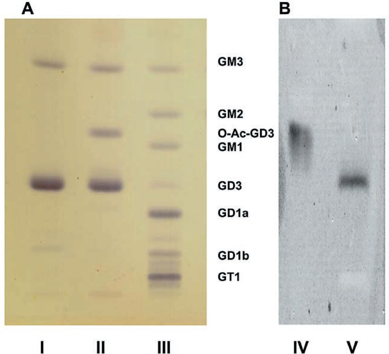
Fig. 1.
Thin-layer chromatogram of brain gangliosides. Technical details are described in the text. A: Stained with the resorcinol spray; B: Stained with specific mouse monoclonal antibodies. Lane I: GalNAcT−/− mouse preparation after saponification; Lane II: GalNAcT−/− mouse preparation before saponification; Lane III: Preparation from Niemann-Pick type C brain as a control (This is only to show thin-layer chromatographic mobility of multiple gangliosides of known structures. The normal control brain used for quantitative analyses of gangliosides (Table I) was a wild-type mouse brain, which is not on this chromatogram.) Lane IV: Same as Lane II but stained with specific monoclonal antibody against O-acetylated GD3; Lane V: Same as Lane II but stained with specific monoclonal antibody against GD3. GalNAcT−/− brain contains essentially GD3, O-acetyl-GD3 and GM3 gangliosides. Upon saponification O-acetyl-GD3 disappears and the GD3 band becomes heavier (see Table I). We tentatively identify the band that runs slightly ahead of GD1b in Lane I as GT3, and the band running slightly ahead of GD1a in Lane II as O-acetylated GT3. Then, the mutant brain also contains detactable amounts of GT3 and O-acetylated GT3, and the latter is hydrolyzed to GT3 by saponificaiton. This interpretation can also be understood from the metabolic block in the GalNAcT−/− mouse.

