. 2014 Oct 23;97(3):439–446. doi: 10.1189/jlb.5HI0414-196R

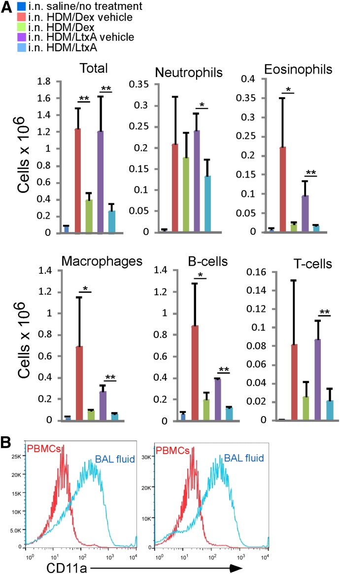
Figure 4. Effects of experimental treatment on WBCs in the BALF in a mouse model for allergic asthma. Saline-treated mice were given saline i.n., whereas all of the other groups were administered HDM extract i.n. HDM extract-exposed mice were then treated with dexamethasone or LtxA or the respective vehicles, as described in the text. (A) Cells in the BALF were stained with mAb for specific cell types and then analyzed by use of flow cytometry. Each bar represents the average from 4 mice. Error bars show sd. The significance of the differences was determined by use of Student’s t-test (*P ≤ 0.05; **P ≤ 0.01). (B) High expression of LFA-1 characterizes cells that have migrated into the airways. PBMCs and BALF WBCs from 2 HDM extract-exposed mice that were treated with LtxA vehicle were stained with anti-CD11a antibody and analyzed by use of flow cytometry.

Figure 4.