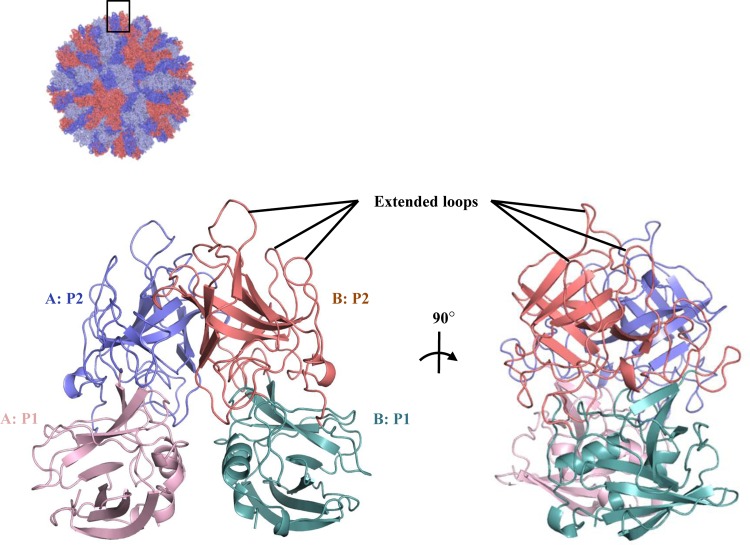
FIG 1.
X-ray crystal structure of unliganded RHDVb P domain. The RHDVa pseudoatomic VLP model (T=3) was modeled with different monomer interactions (12), where each A, B, and C monomer was colored blue, salmon, and light blue, respectively. The boxed region shows the location of the P dimer. The unliganded RHDVb P domain dimer was colored according to monomers (chain A and B) and P1 and P2 subdomains, i.e., chain A, P1, light pink; chain A, P2, slate; chain B, P1, light teal; and chain B, P2, deep salmon. Three extended loops were located on the outer region of the P domain.

