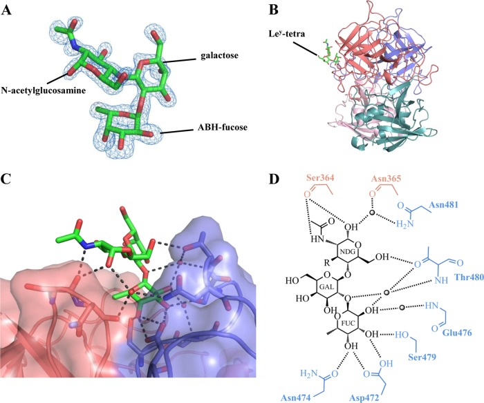
FIG 4.
RHDVb P domain and Ley-tetra interactions. The Ley-tetra is an α-l-fucose-(1-2)-β-d-galactose-(1-4)-N-acetyl-β-d-glucosamine-(3-1)-α-l-fucose. The reducing-end hydroxyl group was fixed in the α position in the Ley-tetra (underlined). (A) The Fo-Fc simulated annealing difference omit map (blue) was contoured at 3.0 σ. The ABH-fucose, galactose, and N-acetylglucosamine (green sticks) of Ley-tetra easily fit into the electron density. The Lewis-fucose was disordered and was not modeled into the structure. (B) The Ley-tetra bound to the dimeric interface on the side of the P dimer (colored as described in the legend to Fig. 1). (C) Closeup surface and ribbon representation of the RHDVb P dimer showing the bound Ley-tetra. (D) The Ley-tetra binding interaction with RHDVb P dimer, where the black dotted lines represent the hydrogen bonds and the sphere represents water molecules (fucose, FUC; galactose, GAL; N-acetylglucosamine, NDG). The Lewis-fucose was disordered and was not modeled into the structure (-R). For simplicity, only the backbone is shown for residues that were backbone mediated. Hydrogen bond distances were less than 3.2 Å, although the majority were ∼2.8 Å.

