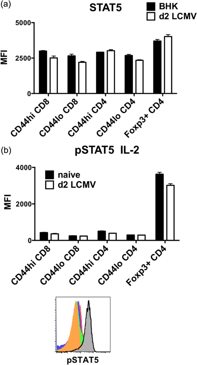
FIG 7.

Foxp3+ CD4+ Treg cells display the highest level of STAT5 expression and respond well to IL-2 stimulation in vitro even after LCMV infection. Foxp3-GFP mice were infected i.p. with 5 × 10⁴ PFU LCMV. (a) Splenocytes harvested at 2 days postinfection were stained ex vivo for STAT5. (b) Phosphorylation of STAT5 (pSTAT5) was assessed among CD44hi CD8 (solid red), CD44lo CD8 (solid blue), CD44hi Foxp3-negative CD4 (solid green), CD44lo Foxp3-negative CD4 (solid orange), and Foxp3+ CD4 (solid gray) T cells from a representative naive animal and Foxp3+ CD4 T cells (black line) from a day 2 LCMV-infected animal after stimulating splenocytes with 5 ng/ml IL-2 for 15 min at 37°C. Data are representative of two separate experiments showing geometric mean fluorescence intensity between BHK sham control or naive versus day 2 LCMV-infected groups of 2 or 3 mice.
