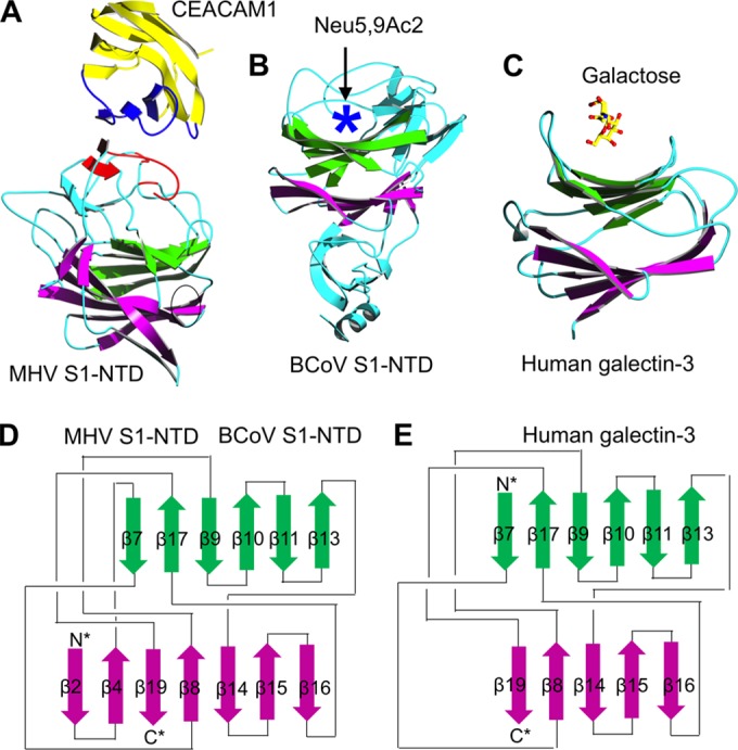
FIG 5.

Crystal structures of coronavirus S1-NTDs. (A and B) These structures include β-genus MHV S1-NTD complexed with its receptor CEACAM1 (PDB ID: 3R4D) (81) (A) and β-genus BCoV S1-NTD by itself (PDB ID: 4H14) (53) (B). (C) The structure of human galectin-3 (PDB ID: 1A3K) is shown as a comparison. In the structure of the MHV S1-NTD-CEACAM1 complex, the VBM on CEACAM1 and the RBM on MHV S1-NTD are in blue and red, respectively. In the structure of BCoV S1-NTD, the sugar-binding pocket as identified by mutagenesis studies is indicated by a five-pointed star. (D and E) The structural topologies of the two coronavirus S1-NTDs (D) and human galectin-3 (E) are shown as schematic illustrations, where β-strands are depicted as arrows and α-helices as cylinders. In the structural topologies of these proteins, the secondary structures are colored and numbered in the same way as for the MHV S1-NTD.
