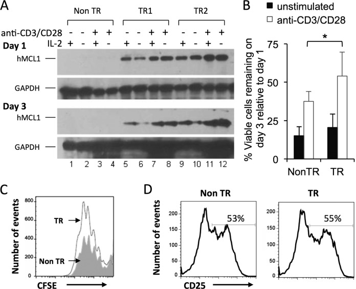
FIG 1.
Anti-CD3 and anti-CD28 antibodies increase MCL1 transgene expression in splenocytes, prolonging their survival in culture. Splenocytes from MCL1 transgenic (TR) and nontransgenic (Non TR) mice were explanted into tissue culture and exposed to anti-CD3 and anti-CD28 antibodies. (A) Transgene expression was monitored (Western blotting) on days 1 and 3 in the absence or presence of recombinant murine IL-2. The lack of signal in cells from nontransgenic animals (lanes 1 to 4) was consistent with the fact that human (but not mouse) MCL1 is detected by the antibodies under the conditions used. (B) Viable cell numbers were monitored by 7-AAD staining. The percentage of viable cells remaining on day 3, relative to day 1, is shown, and represents the mean value of eight independent pairs of transgenic and nontransgenic animals. *, P < 0.05 (for the difference between transgenic and nontransgenic cultures; ANOVA). (C) Cell proliferation (CFSE) was examined 3 days after the application of anti-CD3 and anti-CD28 antibodies. The filled gray histogram represents nontransgenic splenocytes, and the open histogram represents MCL1 transgenic splenocytes, in the 7AAD− (viable-cell) gate. (D) Expression of the T cell activation marker CD25 was examined on day 1.

