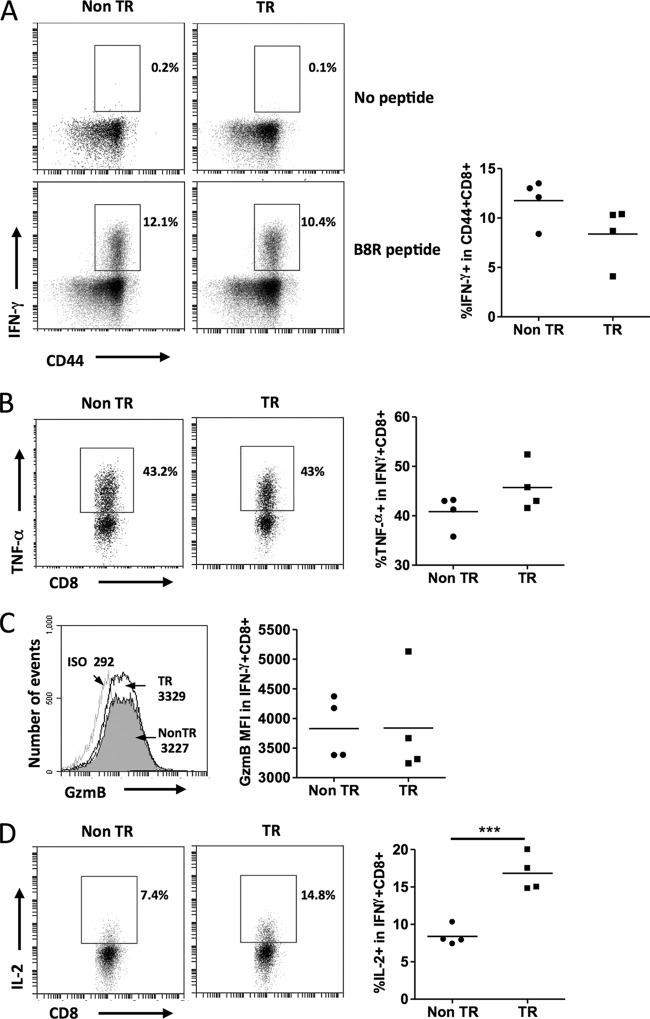
FIG 4.
Effector function during primary viral infection remains intact in the presence of the MCL1 transgene. Mice were infected with VV-WR, and their spleens were harvested at day 10 postinfection. Splenocytes were restimulated with or without B8R peptide for 5 h in the presence of brefeldin A. Intracellular staining was performed to determine IFN-γ production by CD44+ CD8+ T cells (A). No peptide stimulation is shown as a negative control. The percentages in the representative fluorescence-activated cell sorter plots and graphs are those within the CD44+ CD8+ gate. (B) Percentages of IFN-γ+ CD8+ cells that were TNF-α+. (C) Granzyme B (GzmB) mean fluorescence intensity in IFN-γ+ CD8+ cells (filled, nontransgenic [Non TR]; open, transgenic [TR]). Antibody isotype (ISO) for GzmB (light gray line) was used as a negative control for staining. (D) Percentages of IFN-γ+ CD8+ cells that were IL-2+. The data shown are representative of two independent experiments with four mice per experiment. ***, P < 0.0001 (for the difference between transgenic and nontransgenic animals; Student's t test).

