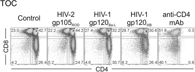
FIG 5.

The X4-tropic HIV-2 envelope per se does not impact the thymocyte distribution in TOCs. TOCs were cultured for 7 days with medium only (control) or in the presence of the recombinant envelope protein HIV-2 gp105ROD, HIV-1 gp120Ba-L, or HIV-1 gp120IIIB (all at 1 μg/ml) or anti-CD4 MAb. The dot plots show a representative example (from one of three independent experiments with different thymuses) of the frequency of subsets in TOCs, as determined by CD4 and CD8 expression. Numbers correspond to the proportion of cells inside gates.
