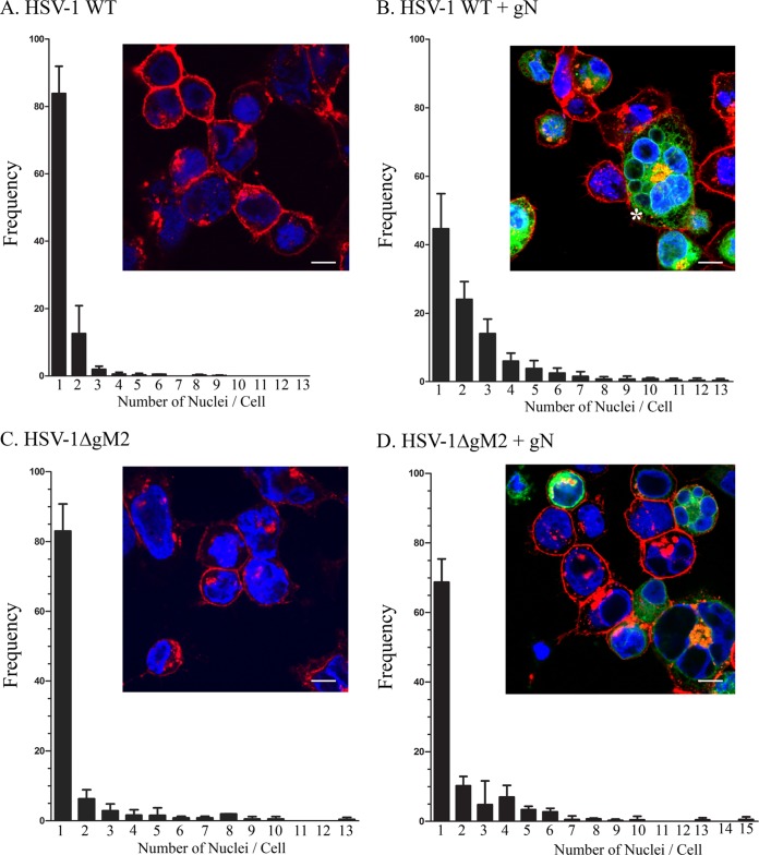
FIG 7.
The HSV-1 gN viral protein induces syncytial formation in infected cells. 143B cells were transfected with (B and D) or without (A and C) YFP-tagged gN for 24 h and subsequently infected at an MOI of 2 with wild-type virus (A and B) or the HSV-1ΔgM2 mutant (C and D). At 12 hpi, the cells were fixed and stained without permeabilization for plasma membrane with Alexa 647-labeled wheat germ agglutinin to delineate the cell boundaries. Cells were then washed and examined by confocal laser scanning microscopy for the presence of syncytia, defined as single cells containing two or more nuclei. Quantification was done by counting 200 cells for each experiment. The reported values represent averages from three independent experiments. The error bars indicate standard deviations. Fluorescence microscopy insets show typical examples for each condition. The asterisk in panel B denotes an example of a syncytium. Scale bar, 10 μm.

