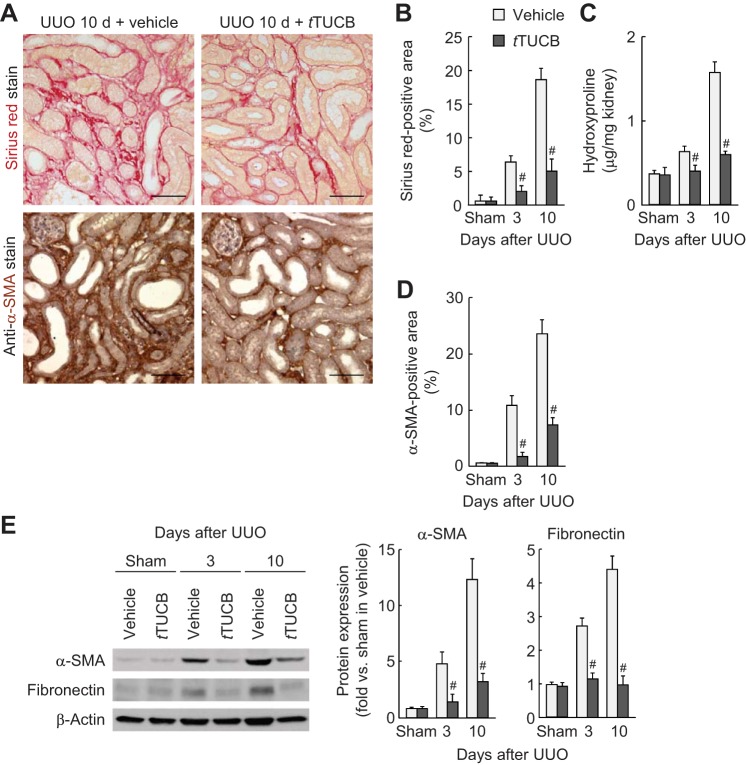
Fig. 2.
sEH inhibition reduces interstitial fibrogenesis during UUO. A: collagen deposition detected by Sirius red stain and α-smooth muscle actin (α-SMA) expression stained with α-SMA monoclonal antibody on kidney sections in vehicle- or t-TUCB-treated mice at 10 days after sham operation or UUO. Scale bars = 50 μm. B: percentage of Sirius red-positive areas on kidney sections. C: collagen content represented by hydroxyproline level in kidneys. D: percentage of α-SMA-positive areas on kidney sections. E: profibrotic expression of α-SMA and fibronectin in kidneys using Western blot analysis. Bands were quantified using Lab Works analysis software. Values are means ± SD; n = 6. #P < 0.05 vs. vehicle.

