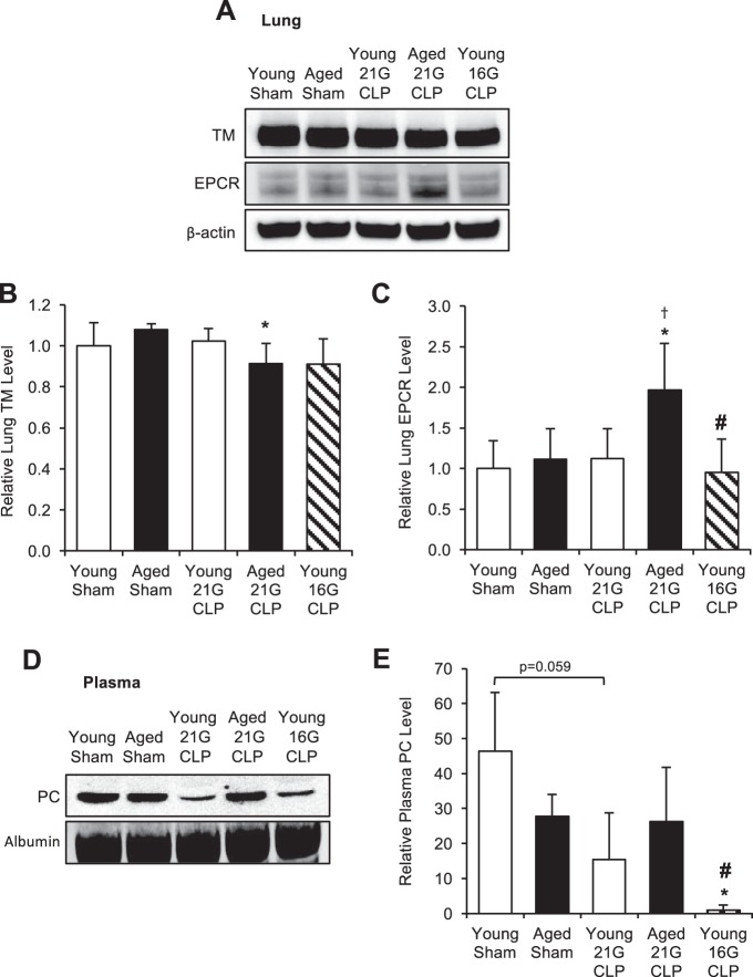
Fig. 5.
Change in levels of protein C (PC) pathway components during sepsis. Young and aged male C57BL/6 mice received CLP surgery with a 16G or 21G needle. Age-matched sham-operated mice were used as controls. A–C: thrombomodulin (TM) and endothelial protein C receptor (EPCR) levels in the lung 24 h after surgery were assessed by Western blot analysis (pooled blot shown) and quantified by densitometric analysis of individually run protein samples. Each lane shown represents a pooled protein sample derived equally from 4 to 5 mice. Membranes were reprobed for β-actin to assure equal protein loading, and quantified levels were adjusted for β-actin. This analysis was performed twice in different sets of mice with the same result. D and E: PC level in the plasma 24 h after surgery was assessed by Western blot analysis (pooled blot shown) and quantified by densitometric analysis of individually run protein samples. This analysis was performed twice in different sets of mice with the same result. Values are means ± SD; n = 4 to 5. *Statistical significance compared with sham-operated mice of the same age; †statistical significance compared with young mice with 21G CLP; #statistical significance compared with aged mice with 21G CLP. One symbol signifies P < 0.05.

