Fig. 7.
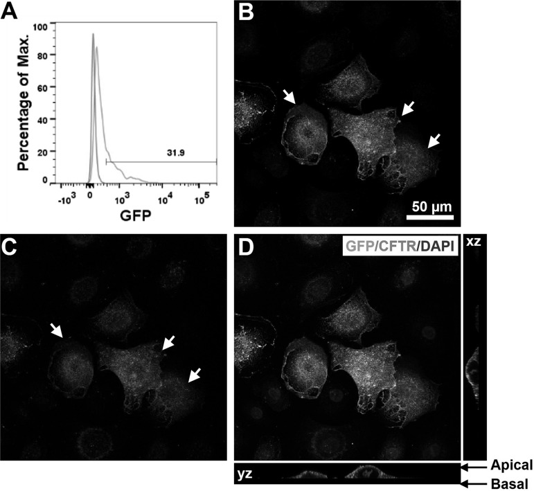
Expression and localization of CFTR-GFP in NHBE cells. A: flow cytometry analysis of NHBE cells infected by pSicoR-CFTR-GFP lentivirus. B–E: confocal images of NHBE cells infected with pSicoR-CFTR-GFP lentivirus. B: GFP fluorescence. C: CFTR immunofluorescence. CFTR was detected with mouse monoclonal anti-CFTR followed by Alexa Fluor 555-conjugated IgG (red). D and E: overlay of GFP fluorescence, CFTR immunofluorescence, and DAPI staining. Arrows, overexpression of CFTR-GFP.

