
Figure 3.
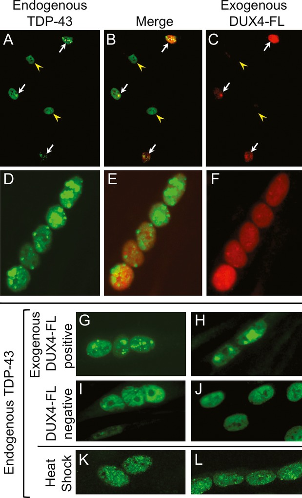
Exogenous DUX4-FL induced aggregation of TDP-43 in myonuclei. (A–C) DUX4-FL (red) was expressed in proliferating human myoblasts using plasmid transfection. Arrows indicate DUX4-FL-positive myoblasts and arrowheads indicate DUX4-FL-negative myoblasts. (D–H) DUX4-FL was expressed in differentiated myotubes using a BacMam vector. Cultures were established with myoblasts from a healthy donor and examined at 32–48 h after addition of plasmids or BacMam vector. TDP-43 immunostaining (green) in DUX4-FL-negative myoblasts (A–C) and myotubes (I and J) was diffusely distributed in nuclei and absent from the cytoplasm, whereas DUX4-FL-positive myoblasts (A–C) and myotubes (D–H) showed a different, punctate pattern of TDP-43 staining indicative of aggregation. (K and L) TDP-43 immunostaining also showed a punctate pattern after a short heat shock (1 h at 42°C).