
Figure 4.
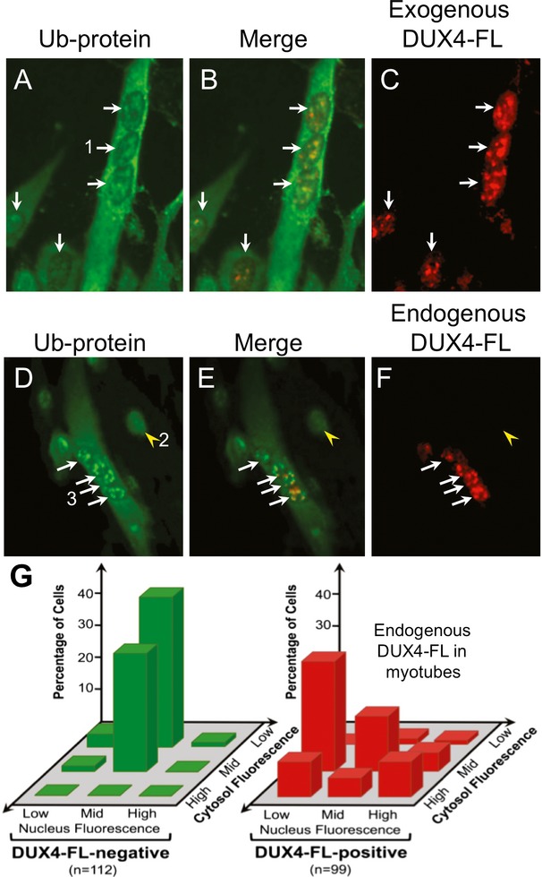
Exogenous and endogenous DUX4-FL altered ubiquitinated protein distribution in myotubes. (A–C) A BacMam vector was used to express exogenous DUX4-FL (red) in myotubes. When compared to the low cytoplasmic and uniform nuclear staining for Ub-proteins in DUX4-FL-negative cells (e.g. Fig.2A and arrowhead in D), DUX4-FL-positive myotubes show increased immunostaining for Ub-proteins (green) in the cytoplasm and variable staining, often with aggregates, in nuclei. (D–F) Expression of DUX4-FL from its endogenous promoter in myotubes formed from FSHD myoblasts (17Abic) also led to increased cytoplasmic staining and variable, often punctate, staining for Ub-proteins. Arrows in all panels indicate cells with DUX4-FL-positive nuclei and the arrowheads in (D–F) indicate a cell with a DUX4-FL-negative nucleus. Additional examples of Ub-protein staining in DUX4-FL-negative cells are shown in Figure7E and F. Numbered cells give examples of the classifications of Ub-protein staining patterns graphed in (G); with the (DUX4-FL-positive) myotube labeled “1” in (A) classified as mid nuclear and high cytoplasmic; the (DUX4-FL-negative) myocyte labeled “2” in (D) classified as mid nuclear and low cytoplasmic; and the (DUX4-FL-positive) myotube labeled “3” in (D) classified as high nuclear and mid cytoplasmic. Note that mid and high-nuclear staining can be punctate, rather than uniform across the nuclei. Additional examples are in Figure2. (G) Nuclear and cytoplasmic Ub-protein staining patterns were independently classified as low, mid, or high, and the number of cells with each pattern was counted. The results showed that endogenous DUX4-FL expression in myotubes increased cytoplasmic staining and produced variable changes in nuclear staining for Ub-proteins.