
Figure 8.
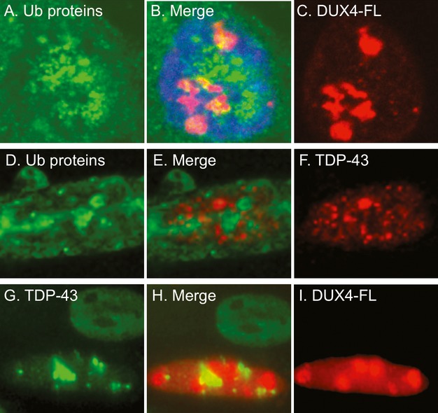
The punctate immunostaining patterns for ubiquitinated proteins, TDP-43, and DUX4-FL in nuclei showed little co-localization. (A–C) Confocal microscope images for ubiquitinated proteins (green, A) and DUX4-FL (red, C) were merged in (B) to show the relatively small regions of overlap (yellow). (B) Includes blue DNA stain for the nucleus. (D–F) Standard microscope images for ubiquitinated proteins (green, D) and TDP-43 (red, F) were merged in (E) and showed little overlap of the punctate areas of immunostaining in the nucleus. (G–I) Standard microscope images for TDP-43 (green, G) and DUX4-FL (red, I) were merged in (H) and showed little overlap of the punctate areas immunostaining seen in the lower DUX4-FL-positive nucleus. Additional examples are in Figure5. The upper DUX4-FL-negative nucleus in (G–I) shows the nearly uniform nuclear immunostaining for TDP-43 that is seen in the absence of DUX4-FL.