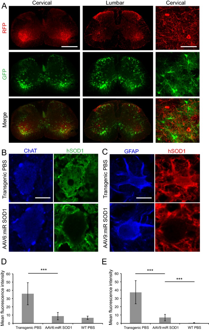
Figure 3.

SOD1 expression is suppressed in motoneurons and astrocytes of end-stage animals injected with AAV-miR SOD1. (A) RFP and GFP expression in motoneurons or astrocytes, respectively, is conserved in end-stage animals injected with various vector combinations. Represented are cervical and lumbar sections of a mouse from the AAV6 + AAV9:miR SOD1 group. Scale bar: whole spinal cord, 500 μm; cervical close-up, 80 μm. (B) Human SOD1 expression in a motoneuron of a PBS-injected transgenic mouse as compared to an AAV6-cmv:RFP:miR SOD1 injected animal. Note the absence of human SOD1 labeling in the vector-injected condition. Scale bar: 15 μm. (C) Human SOD1 expression in astrocytes of a transgenic mouse injected with PBS or AAV9-gfaABC1D:GFP:miR SOD1. In the latter group, note the loss of human SOD1 expression in the cell body of the GFAP-labeled astrocyte. Scale bar: 10 μm. (D) Immunolabeled human SOD1 level expressed as mean fluorescence intensity. Mean fluorescence intensity was randomly assessed in 20 motoneurons identified by ChAT immunostaining for each represented condition. Human SOD1 levels are significantly lower in end-stage animals injected with AAV6-cmv:RFP:miR SOD1 as compared to PBS-injected transgenic mice. (E) Mean fluorescence intensity following human SOD1 immunostaining in astrocytes of AAV9:miR SOD1, transgenic PBS or WT PBS groups. Note the silencing of human SOD1 expression in animals injected with AAV9-gfaABC1D:GFP:miR SOD1. N = 20 motoneurons or astrocytes per animal, three animals per condition. ***P < 0.001, one-way ANOVA and Newman–Keuls post hoc test. Data are represented as mean ± standard deviation. SOD1, superoxide dismutase 1; WT, wild-type.
