Figure 4.
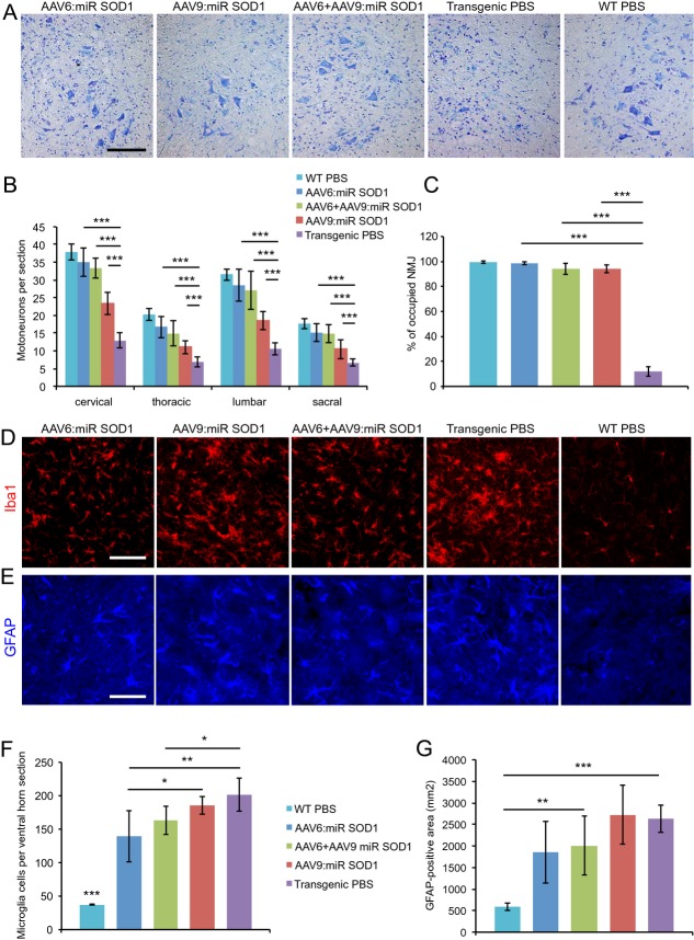
Motoneuron counts and neuromuscular junction occupancy are significantly increased in all treated groups despite astrogliosis and microglial activation. (A) Cresyl violet staining of lumbar ventral horns showing motoneuron preservation despite inflammatory infiltration for all treated conditions. Scale bar: 100 μm. (B) Motoneuron counts per spinal cord section for vector-injected and control animals at cervical, thoracic, lumbar, and sacral levels. Motoneuron numbers are significantly increased at all levels in treated animals. AAV6:miR SOD1: n = 9, AAV6 + AAV9:miR SOD1: n = 7, AAV9:miR SOD1: n = 8, transgenic PBS: n = 12, WT PBS: n = 14. (C) Neuromuscular junction integrity expressed as percentage of SV-2-positive motoneuron terminals to bungarotoxin-positive motor end plates. Innervation is preserved in all vector-injected conditions (n = 3 per group). (D) Microglial activation evidenced by increased Iba1 immunostaining in transgenic animals. Scale bar: 100 μm. (E) GFAP immunostaining showing astroglial activation. (F) Microglial counts per lumbar ventral horn in vector-injected and control mice. Note that AAV6:miR SOD1 and AAV6 + AAV9:miR SOD1 conditions display significantly decreased microglial activation compared to transgenic PBS and AAV9:miR SOD1 groups; n = 5 animals per group, 10 ventral horns per animal. (G) Astrogliosis determined by measuring GFAP-positive total area in lumbar ventral horns of vector-injected and control animals; n = 5 animals per group, 10 ventral horns per animal. Data are expressed as mean ± standard deviation. *P < 0.05, **P < 0.01, ***P < 0.001, one-way ANOVA and Newman–Keuls post hoc test. SOD1, superoxide dismutase 1.

