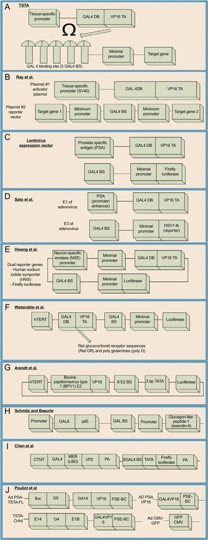
Figure 4.3. See legend on next page.
Schematic diagrams of various TSTA approaches. (A) Original TSTA approach. A weak, tissue-specific promoter is used to drive the expression of a fusion protein that will bind to its binding sites and amplify the expression of a target gene. (B) Bidirectional vector with two target genes of interest that would be amplified when bound to the chi-meric protein created as a result of the tissue-specific promoter SV40. (C) Lenti-TSTA. The TSTA was inserted into a lentivirus expression system with the PSA promoter to amplify expression of a reporter gene such as fLuc. (D) Adeno-TSTA. The system was optimized to utilize the E1 and E3 segments of the adenovirus to enhance expression of the reporter gene HSV1-tk using PSA acting as a promoter for synthesis of the chimeric protein. (E) TSTA system for imaging cellular processes such as neuronal differentiation via the NSE promoter. (F) Addition of rat glucocorticoid receptor sequences (Rat GR) and poly-glutamines (polyG)in between the GAL4 and VP16 domains to increase efficiency of luciferase expression. (G) Replacing GAL4-UAS with a bovine papillomavirus 1 transcriptional activator. (H) Replacing VP-16 with p65. (I) Titrable TSTA using the cardiac troponin T (CTNT) promoter and the MER sequence responsive to raloxifene. (J) Dual TSTA system (dTSTA) for the expression of a reporter and a conditionally replicating Ad.

