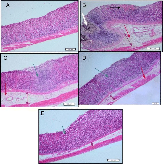
Figure 7.

Histological evaluation of gastric lesions in sections stained with Hematoxylin & Eosin (10X). Normal control group (A) has normal tissue epithelium. Indomethacin group (B) shows disruption of surface epithelium with hemorrhage (black arrow) and the lesions infiltrate deep into mucosa layer (white arrow) wide edema (brown arrow) and leukocyte infiltration (red arrow). HPTP pre-treated groups (C & D) shows reduction of submucosal edema (brown arrow), mild to moderate disruption of mucosa epithelium (blue arrow) few infiltration of leukocyte. Omeprazole group (E) shows mild disruption of the surface epithelium (blue arrow), and mild submucosal edema (brown arrow) and bit of leukocyte (H&E stain, magnification 20x).
