
Fig. 1.
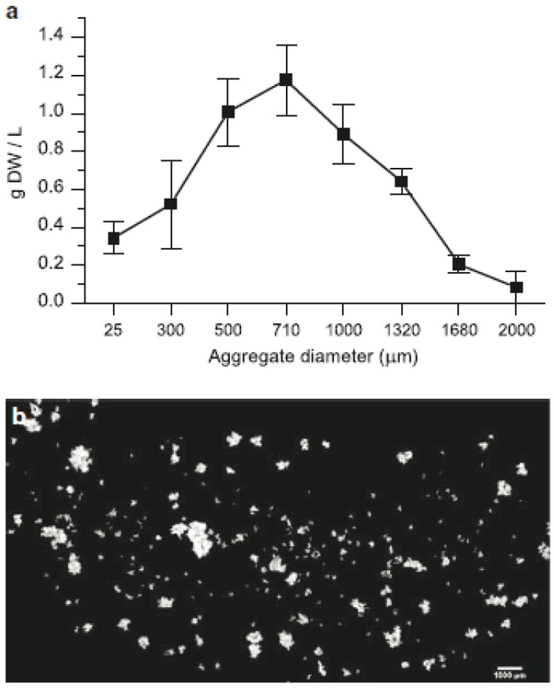
Typical size and morphology of aggregates in Taxus cuspidata suspension culture. a Distribution obtained from filtration of cultures on day 7; data points represent biomass retained on corresponding filter size, error bars represent standard deviations from three replicate flasks. b Composite image of aggregates stained with flourescein diacetate