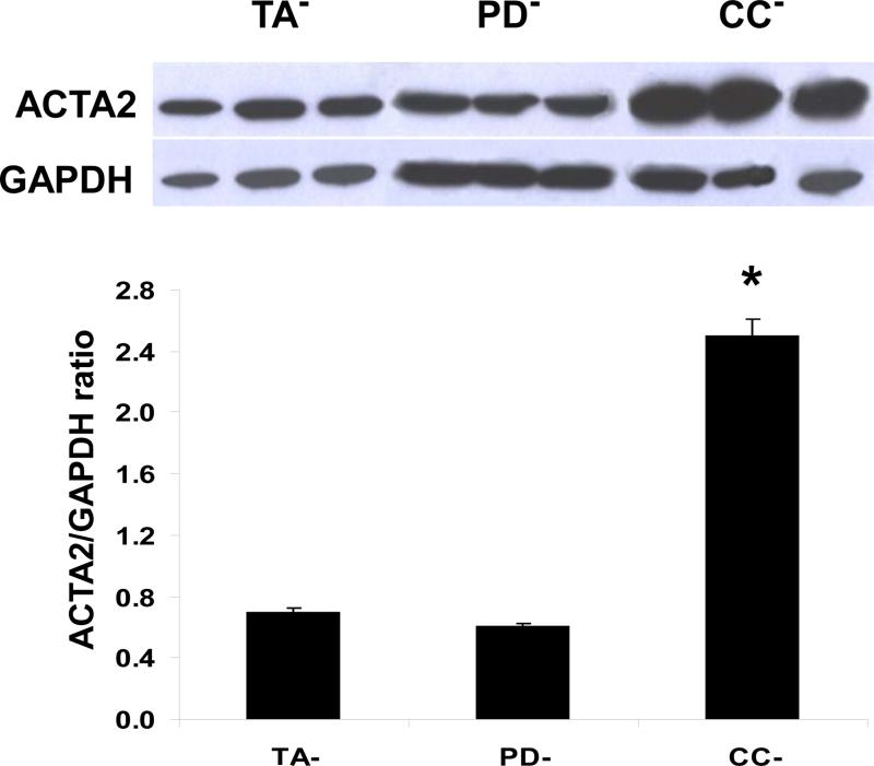
Figure 1. The content of activated myofibroblasts in the non-stimulated PD culture (PD−) was not higher than in the non-stimulated non-PD tunica albuginea culture (TA−), as shown by the expression of ACTA2 protein.
PD− and TA− cell cultures maintained in regular DMEM/20% fetal calf serum for 8-15 passages in the absence of added TGFβ1 were essentially fibroblast cultures, as assayed with vimentin, whereas CC− cultures were essentially SMC, as assayed with calponin (24-26). Cells on monolayer in 6-well plates were homogenized from triplicate wells for each culture and subjected to quantitative western blot assay for ACTA2 expression, correcting by GAPDH expression. In the case of the CC− cultures, ACTA2 does not discriminate myofibroblasts from SMC. *p<0.05

