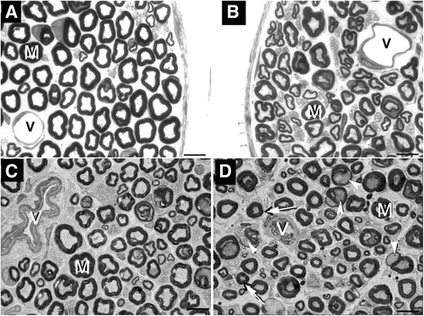
Figure 1.

Representative semithin cross section of the sural nerve from normotensive Wistar rat. (A), normotensive Wistar rat with chronically induced diabetes (B), Spontaneously hypertensive rat (C) and Spontaneously hypertensive rat with chronically induced diabetes (D), showing typical endoneural structures. Large (M) and small myelinated fibers are present in the endoneural space. Note the presence of normal endoneural vessels (V) in A and B while in C and D, the vessels show thickening of the walls and reduced lumen. In D, arrows indicate axons with atrophy and arrowheads indicate myelinated fibers with severe myelin disruption. Toluidine blue stained. Bar = 10 μm.
