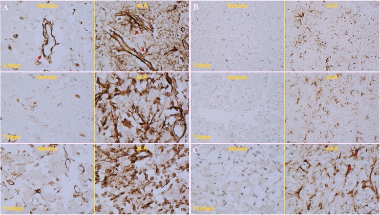
Figure 7.

IHC staining for anti-nestin (A) and GFAP (B, C) protein following ischemia in the infarct core region at 3, 7, and 14 days after ischemia (magnification, A, C 400×; B 200×). Compared with the corresponding regions in the control group, there was an increase in nestin- and GFAP-positive cells and vessels in the aLA group at 3, 7, and 14 days after the MCAO. Additionally, the morphological features of nestin- and GFAP-expressing cells and vessels, such as the number and shape of cell bodies and processes, were significantly improved in the aLA group (arrow and star). In contrast, the control group exhibited loss of processes (arrowhead). n = 3 rats/day/group.
