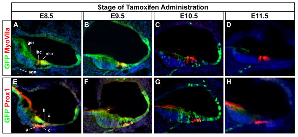
Figure 3. Hair and support cells in the organ of Corti derive from Wnt responsive progenitors.

Transverse sections through the cochlear duct of TopCreER; RosaZsGreen/+ embryos (E18.5) exposed to tamoxifen at different developmental stages. (A–D) Co-labeling of the GFP+ (green) Wnt responsive lineage with MyoVIIa+ (red) hair cells. Inner and outer hair cells (ihc, ohc) showed co-labeling (yellow) when tamoxifen was administered between E8.5 and E9.5, but not thereafter. (E–H) Co-labeling of GFP (green) and Prox1 (red) a support cell marker that labels pillar (p) and Dieters’ (d) cells. Support cells showed co-labeling (yellow) when tamoxifen was administered between E8.5 and E10.5, but not thereafter. GFP staining was also detected in cell types adjacent to the organ of Corti, including the greater epithelial ridge (ger), Hensen’s cells (h), Claudius’ cells (c), and spiral ganglion neurons (sgn).
