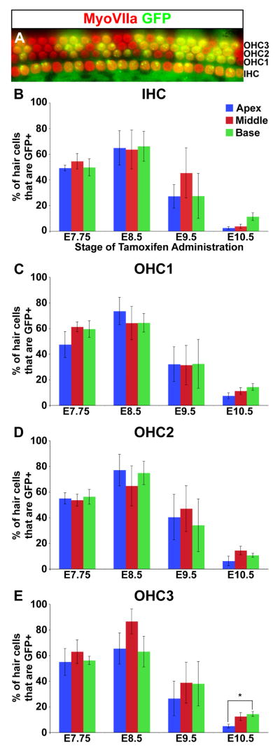
Figure 4. TopCreER marks hair cells along the entire length of the cochlear duct.
(A) An optical section through the cochlear sensory epithelium of an E18.5 TopCreER; RosaZsGreen/+ embryo exposed to tamoxifen at E8.5. The cochlea was immunostained with an antibody against MyoVIIa (red). GFP (green) was visualized by direct fluorescence. The majority of hair cells show co-labeling (yellow). (B–E) Bar graphs displaying the percentage of TopCreER labeled hair cells at distinct positions along the cochlear duct upon tamoxifen administration between E7.75 and E10.5. The maximum number of TopCreER labeled inner and outer hair cells was observed when tamoxifen was administered at E8.5. The third row of outer hair cells showed a significant reduction in labeling at the apex compared to mid and basal levels when exposed to tamoxifen at E10.5 (p<0.01). Error bars represent s.e.m.

