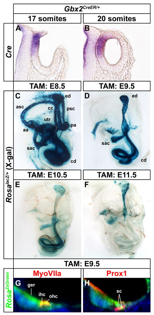
Figure 5. Gbx2CreER/+ labels cells contributing to both vestibular and auditory structures.
(A, B) Transverse sections of Gbx2CreER/+ embryos at otic cup (17 somite) and otic vesicle (20 somite) stages showing dorsomedial localization of Cre mRNA expression. (C–F) Whole mount views of X-gal stained inner ears (E18.5) exposed to tamoxifen at indicated developmental time points to induce Gbx2CreER dependent activation of RosalacZ. Tamoxifen administration between E8.5 and E10.5 labels vestibular and auditory structures, whereas after E11.5, labeling is restricted to the endolymphatic duct. (G, H) Transverse sections through the cochlear duct of Gbx2CreER/+; RosaZsGreen/+ embryos (E18.5) exposed to tamoxifen at E9.5 showing GFP co-labeling with MyoVIIa+ hair cells and Prox1+ support cells. Abbreviations: anterior semicircular canal, asc; posterior semicircular canal, psc; anterior ampulla, aa; posterior ampulla, pa; endolymphatic duct, ed; cochlear duct, cd; utricle, utr; saccule, sac; greater epithelial ridge, ger; inner hair cell, ihc; outer hair cells, ohc; support cells, sc.

