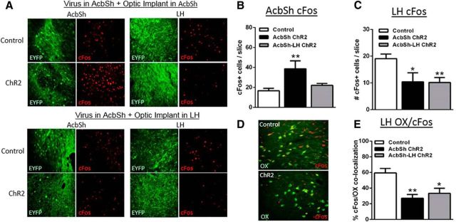
Figure 5.
Optogenetic stimulation of either global AcbSh neurons or selective AcbSh-LH terminals reduces cFos in LH orexin neurons. A, Representative images (40 μm, 20×) of EYFP (green) and cFos (red) immunoreactivity in the AcbSh and LH 90 min after optogenetic stimulation and FST behavioral testing in global AcbSh (top) and selective AcbSh-LH (bottom) stimulated groups. B, AcbSh cFos was elevated only after global AcbSh stimulation. C, LH cFos was reduced after either global AcbSh or selective AcbSh-LH terminal stimulation. D, Representative images showing reductions in cFos (red)/orexin (OX; green) colocalization after optogenetic stimulation. E, cFos/OX colocalization was reduced by either global AcbSh neuron or selective AcbSh-LH terminal stimulation; *p < 0.05, **p < 0.01 versus control; n = 4–9/group.

