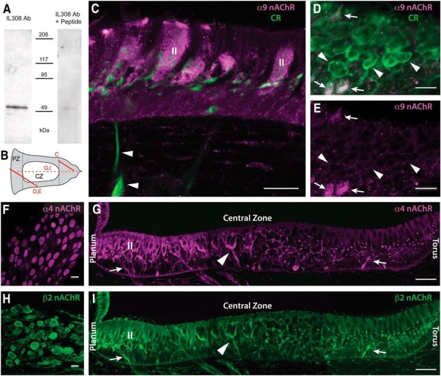
Figure 10.
Immunohistochemistry for α9, α4, and β2 nAChR subunits. A, Western blot with our IL-308 antibody shows that α9 nAChR subunit protein is present as a single band of the predicted molecular weight (≈56 kDa) in turtle inner ear tissue. Preincubation with an excess of antigen peptide (IL 308 Ab + peptide) blocked detection of the band. B, Schematic of turtle hemicrista indicates the plane of sectioning for subsequent panels (Fig. 1, top). C, Antibodies to calretinin (CR) stained bouton afferent nerve fibers (green, arrowheads) innervating type II hair cells in the PZ near the torus. IL-308 stained torus type II hair cells (magenta, II), but not associated bouton endings (green). D, E, Section through the CZ and the adjacent PZ stained for CR and α9 nAChR subunit. Calyx fibers and endings in CZ (D, green, arrowheads) are, at most, very lightly stained with the α9 antibody (E, magenta), while two type II hair cells in adjacent PZ (D, white; E, magenta, arrows) are stained with both CR and the α9 antibodies. F, H, Antibodies to α4 nAChR subunits (F) and β2 nAChR subunits (H) label cells in vestibular ganglia. G, I, Antibodies to α4 and β2 nAChR subunits both label bouton afferents near the torus and planum (arrows), calyx-bearing afferents in the CZ (arrowheads), and type II hair cells (II) near the planum. Both panels are from the same crista section. Scale bars, 20 μm.

