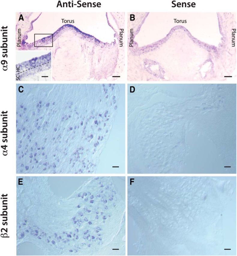
Figure 11.

Expression of α9, α4, and β2 nAChR subunit mRNA in turtle tissue. A, B, C, E, ISH using nAChR subunit-specific antisense probes in the turtle crista (A, B) and vestibular ganglia (C, E). A, In longitudinal sections of the posterior crista (Fig. 10B, diagram), α9 nAChR subunit mRNA was localized to the hair cell layer (HC) with more intense labeling near the torus. Inset, Higher-magnification image of labeled hair cells taken from box. Dashed line demarcates HC and supporting cell (SC) layers. C, E, Both α4 and β2 nAChR subunit mRNA were found in vestibular ganglia, respectively. B, D, F, Sense controls for each nAChR subunit showed no labeling. Scale bars: inset, 20 μm; A, B, 50 μm; C–F, 20 μm.
