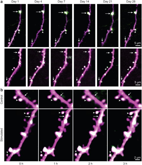
Figure 2. Acute whisker stimulation leads to an increase in spine sGluA1 in vivo in apical dendrites of layer 2/3 neurons in the barrel cortex.
a, Long-term stable expression of SEP-GluA1 and dsRed2. Same spines were marked with arrows and arrowheads in different imaging sessions. b, Representative time-lapse in vivo 2-photon images of layer 2/3 pyramidal cell apical tuft dendrites taken with no whisker stimulation (Control) or before (Stimulated, hour 0) and after 1-hour-long acute whisker stimulation (Stimulated, hour 1, 2, and 3). Arrowheads mark spines and arrows mark dendritic shafts (SEP-GluA1 in green, dsRed2 in magenta, overlap in white). Images are single plane median filtered images that were up-scaled and contrast enhanced.

