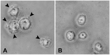
Figure 1.

Efficient release of nuclei from cells using hypo-osmotic buffer and pipetting. Phase-contrast microscopic image of THP-1 cells in Lysis Buffer before (A) and after (B) pipetting through a 200-μl pipette tip. Original magnification: ×200. Arrowheads indicate membrane components around the nuclei that were observed before (A), but not after (B), pipetting.
