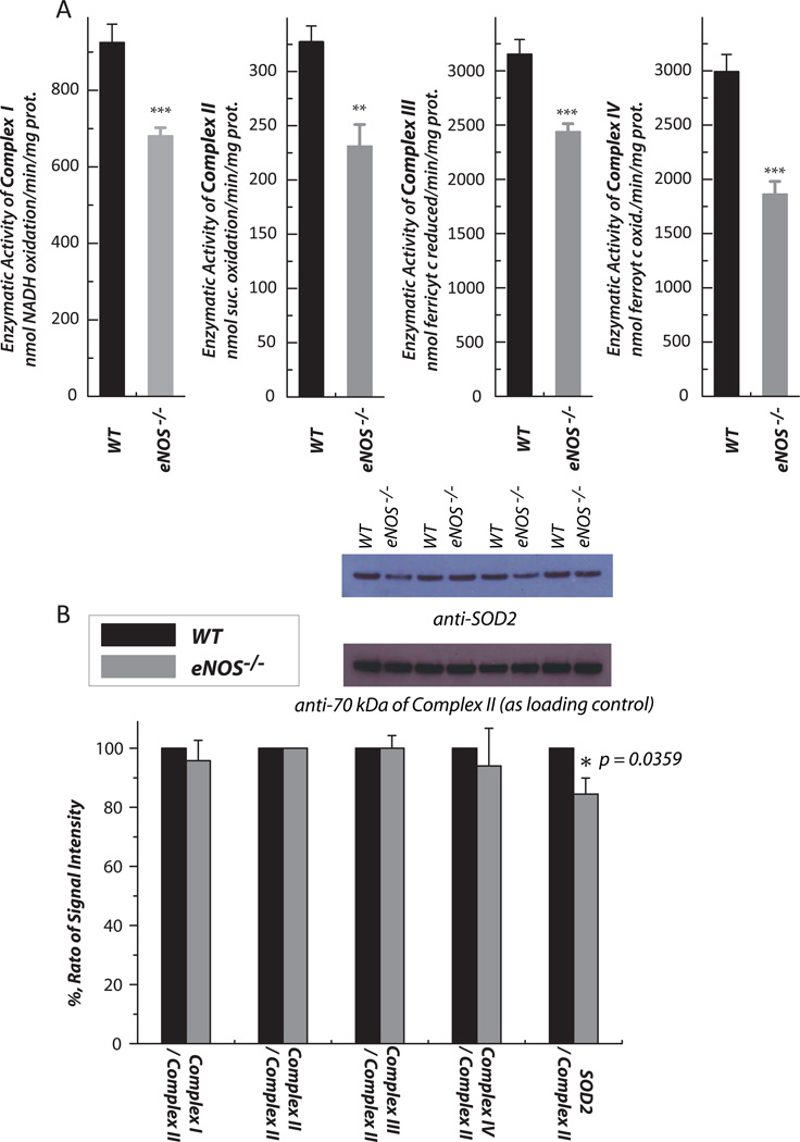
Fig. 4.
Hearts were removed from wild type and eNOS−/− mice, and subjected to mitochondrial preparation. A, enzymatic activities of electron transport chain (ETC) components in the mitochondria were assayed as described previously (n = 7). B, Protein expression of ETC components and SOD2 in the mitochondria was assessed by Western blot using the following antibodies: Ab51 for Complex I (1:10,000)[6], AbGSC90 for Complex II (1:30,000) [54], the monoclonal antibody against Rieske iron-sulfur protein (1:1000) and Cox I (1:3000) for Complexes III and IV, and a polyclonal antibody against SOD2 (1:2000). The protein expression level of subunit I (70 kDa flavoprotein) of Complex II in the mitochondria was used as a loading control. The density ratio of the blotting signals between the ETC components/SOD2 and Complex II was quantitated by software Image J. n = 4; *p < 0.05, **p < 0.01, ***p < 0.001. Data were normalized to the amount (mg) of mitochondrial protein in A.

