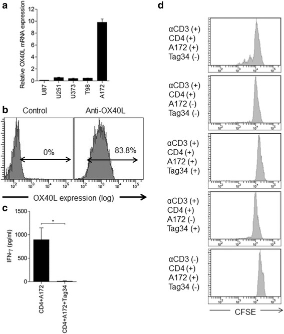
Figure 2.

Endogenous OX40L in human glioblastoma and its functional implication. a. Quantitative PCR of OX40L mRNA expression in five glioblastoma cell lines. b. OX40L protein expression in A172 cells, as determined by flow cytometry. c. Production of IFN-γ from human CD4-positive cells, cocultured with irradiated A172 cells. Tag34, a blocking antibody against human OX40L (*P < 0.05), under anti-CD3 coated plates. d. CFSE staining of human CD4-positive cells detected by flow cytometry. The top panel shows the cell division pattern of CD4-positive cells that were cocultured with irradiated A172 cells in anti-CD3 antibody-coated plates. Compared with the top panel, either the absence of A172 (2nd panel) or the addition of Tag34 (3rd panel), Tag34 only (4th panel) or the absence of anti-CD3 (5th panel), resulted in the loss of the cell division patterns.
