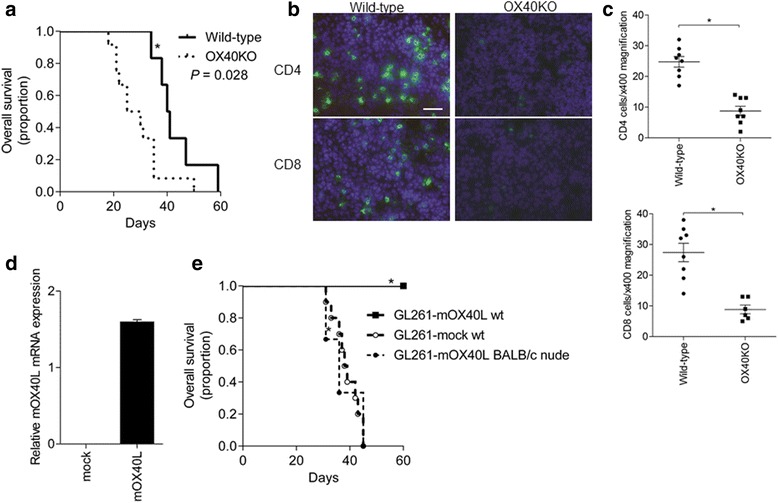
Figure 3.

OX40 signaling in mouse models of glioma. a. Survival curves of OX40KO mice (n = 12) and wild-type mice (n = 6) with intracranial GL261 cells. OX40KO mice bearing the GL261 cell transplant were associated with significantly shorter survival than that of wild-type mice (P = 0.028). b. CD4 and CD8 cells (green) at the sites of the GL261 cell transplants of wild-type and OX40KO mice. Scale bar, 40 μm. c. Numbers of CD4 and CD8 cells, counted under 400× magnification (mean ± SEM) (*P < 0.05). d. Transfection of G261 cells with retrovirus harboring OX40L cDNA or empty vector. The expression of mouse OX40L (mOX40L) was confirmed by quantitative PCR. e. Survival curves. GL261-mOX40L cells were injected into the brain of wild-type mice (wt, n = 10) or BALB/c nude mice (n = 3). GL261-mock cells were injected into the wild-type mouse brain (mock wt, n = 10) for control (*P < 0.0001).
