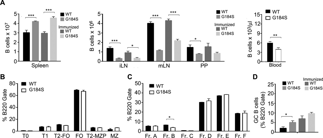
Figure 1.
Altered distribution of B cells in the lymphoid organs of G184S KI bone marrow chimeric mice. (A) Flow cytometric analysis to determine the number of B cells in the spleen, LNs, Peyer’s patches and wild type and G184S KI bone marrow reconstituted mice immunized with sheep RBCs i.p (day 10) or not. Mice were analyzed 8–10 weeks after reconstitution. (B) Flow cytometric analysis of B cells from the spleen of bone marrow chimeric mice to examine splenic B cell development. (C) Flow cytometric analysis of B cells from the bone marrow of chimeric mice to examine B bone B cell development. (D) Flow cytometric analysis of splenic GC B cells from immunized or non-immunized bone marrow reconstituted mice. GC B cells defined as B220+CD38−FAS+GL7+. All experiments independently preformed 3 times with 3~4 mice and presented as the mean ± SEM of 3~4 mice per group.

