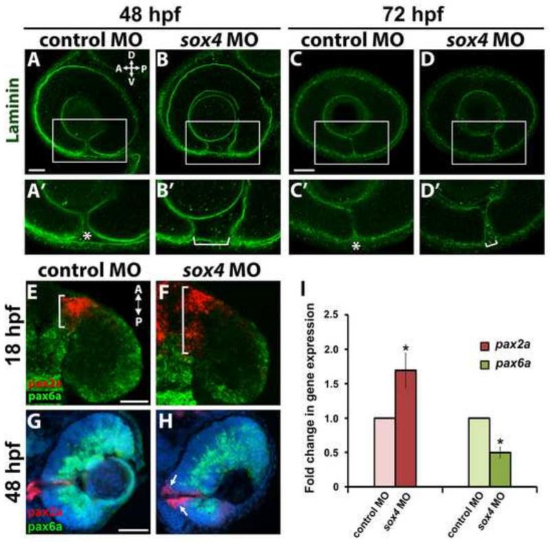
Figure 3. Persistence of laminin expression at the choroid fissure and altered proximo-distal patterning of the optic vesicle in sox4 morphants.
(A-D’) Laminin immunostaining on control and sox4 morphant embryos at 48 and 72 hpf. Representative images from 15-20 individuals analyzed for each group are shown. (E-H) Double FISH for pax2a and pax6a at 18 and 48 hpf. (I) qPCR analysis revealed a 1.69-fold increase in pax2a expression and a 2.0-fold decrease in pax6a expression in sox4 morphant heads at 18 hpf (Student's t-test, P<0.05). All scale bars equal 50 μm. Asterisks in A’ and C’ indicate the closing or fused choroid fissure in control morphants; brackets in B’ and D’ indicate the open choroid fissure in sox4 morphants.

