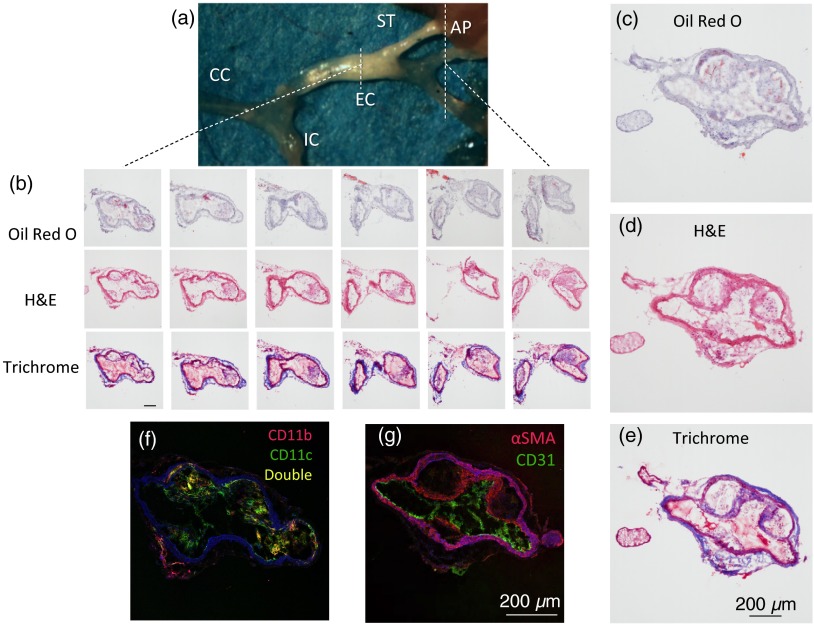
Fig. 2.
(a) Plaque is visible (opaque white) in the external carotid (EC) artery and superior thyroid artery branch of an Apoe−/− mouse fed WD for 12 weeks. Plaques can also develop in the ascending pharyngeal artery and lower EC branches. CC, common carotid artery; IC, internal carotid artery. (b) Serial sections every from an Apoe−/− mouse fed WD 9 weeks were stained for neutral lipids (Oil Red O), cell components (H&E), and collagen and elastin fibers (Trichrome.) . Every other stained section is shown. Larger examples are shown for visibility (c-e). Serial sections were also immunostained for (f) CD11b and CD11c and (g) -smooth muscle actin and CD31. Autofluorescence and nuclei (stained with Yoyo-1) appear in blue. Brightness was increased by 40% on all fluorescence images for visibility.

