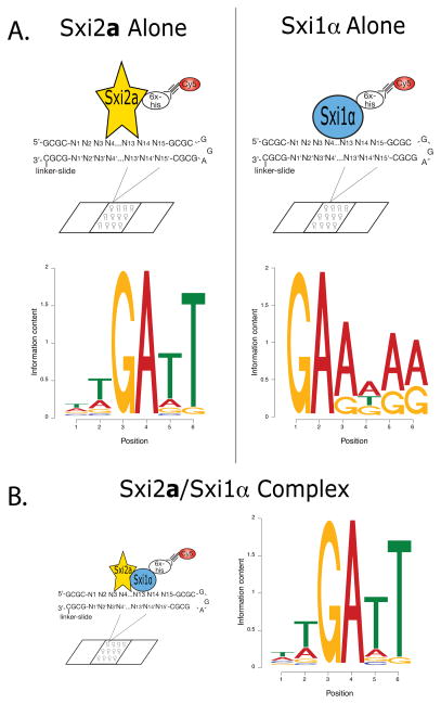
Figure 1. In vitro binding site determination for Sxi2a and Sxi1α.
A. Individual in vitro binding site identification. Full-length Sxi2a (yellow star) and Sxi1α (blue oval) were labeled individually with Cy5 (red circle) and incubated separately with double-stranded 15-mer oligonucleotides spotted on glass slides (CSI arrays). Motifs shown represent the sequences with the highest binding affinities for each protein. B. Heterodimer in vitro binding site identification. Sxi1α was labeled with Cy5, and Sxi2a and Sxi1α were incubated together with the oligonucleotide array. The motif shown represents sequences to which the proteins bound with high relative affinity. In all motifs the height of each individual letter is representative of the conservation of that nucleotide.

