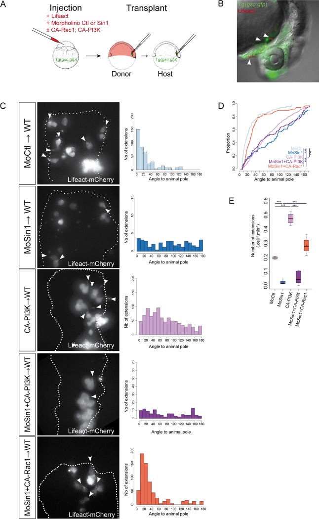
Fig 4. Absence of Sin1 affects protrusive activity of prechordal plate cells.
(A) Diagram of the design of prechordal plate cell transplantation. Cells were labelled with Lifeact-mCherry and transplanted from shield to shield. (B) At 24 hpf, sin1 morphant cells transplanted into wild-type prechordal plates take part to the hatching gland. (C) Cells injected with Lifeact-mCherrry RNAs and either a control morpholino, or the sin1 morpholino, or CA-PI3K RNAs, or the sin1 morpholino and CA-PI3K RNAs, or the sin1 morpholino and CA-Rac1 RNAs were transplanted from shield to shield in Tg(-1.8gsc:GFP) embryos. Transplanted cells are within the prechordal plate (assessed by GFP expression), and contour of the plate is delineated (white dotted line). Orientations of their cytoplasmic extensions were measured relative to the animal pole and plotted as histograms (C) and cumulative plot in (D). (E) Frequencies of cytoplasmic extensions.

