
Fig. 3.
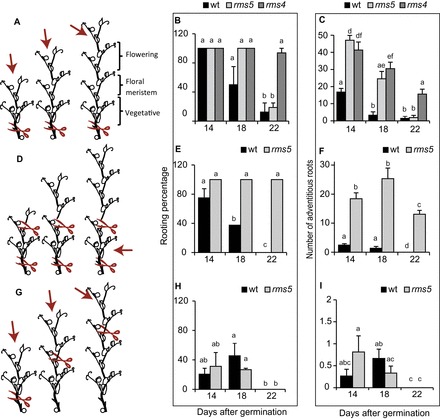
Cuttings can be taken at different chronological ages, or different physiological ages, within or across different ontogenetic ages. (A, D, G) Cartoon representations showing the location of cutting (red scissors) and which section of the stem was used for the adventitious rooting assay (red arrows); however the number of nodes in the older cuttings are not necessarily representative as this changes slightly with genotype (Supplementary Fig. S6). (A) Cuttings taken above node two have different chronological age at the cutting base, different physiological age of the apical meristem, and different size cuttings crossing the vegetative–floral ontogenetic switch. (D) Cuttings taken above node 2 but decapitated have different chronological age at the cutting base, different physiological age of the original apical meristem, and have the same size cuttings crossing the vegetative–floral ontogenetic switch. (G) Cuttings taken with two leaves expanded have bases with the same chronological age, apical bud with different physiological age, and same size cuttings crossing the vegetative–floral ontogenetic switch (Supplementary Table S1). (B, E, H) Changes in rooting percentage with age and (C, F, I) adventitious root formation declines in cuttings taken as illustrated in A, C, and E respectively. Means are presented ± standard error; different letters represent significantly different means (Students t-test). n>16. Rooting percentage was calculated per rooting box and then averaged to produce the standard error bars and statistics. Where bars are missing the standard error was 0. (This figure is available in colour at JXB online.)