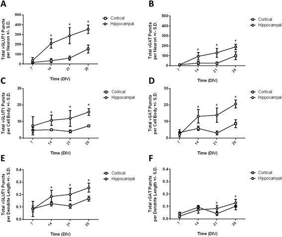
Figure 5.

Excitatory and inhibitory synapse development in hippocampal and cortical neurons as assessed using high content image analysis. Quantification of excitatory (left column) and inhibitory (right column) synaptogenesis in hippocampal (circles) and cortical (squares) using high content image analysis. The number of vGLUT1 or vGAT immunopositive puncta was used to measure the number of excitatory and inhibitory synapses, respectively. (A) Total number of vGLUT1 puncta per neuron. (B) Total number of vGAT puncta per neuron. (C) Total number of vGLUT1 puncta per cell body. (D) Total number of vGAT puncta per cell body. (E) Total number of vGLUT1 puncta per μm dendrite length. (F) Total number of vGAT puncta per μm dendrite length. Data were analyzed by two-way ANOVA. For each endpoint, a significant interaction between time and cell type was observed; therefore, post hoc mean contrast tests were performed to compare means within each cell type across time and means between cell types within each time point. *Means are significantly different between cell types within a time point (Sidak’s test, p < 0.05). All data are expressed as mean ± SD (n = 6–18 wells collected across 3–4 replicate cultures).
