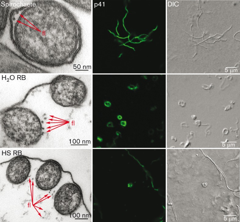
Fig. 6.

Flagella are present in RBs. TEM and confocal micrographs of flagella localization in spirochaetes, 2 h H2O RBs and 4 days HS RBs. TEM images (left panels) demonstrate the cross-section of the cell, where flagella (fl) are indicated by arrows. Middle panels display confocal images of cells immunolabelled with p41 flagellin protein antibody and Alexa 488 (green). Right panels illustrate the DIC image.
