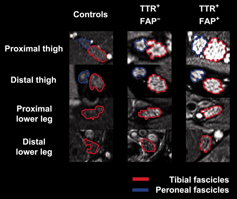
Figure 4.
Magnetic resonance neurography source images. Representative source images (right leg; high-resolution T2-weighted turbo spin echo fat-saturated sequences, 3 T) with overlaid coloured regions of interest indicating segmented tibial and peroneal fascicles within the sciatic nerve (at proximal thigh and distal thigh levels) and the distal continuation of tibial fascicles as tibial nerve (at proximal lower leg and distal lower leg levels). One healthy control, an asymptomatic gene carrier (TTR+-FAP−) and a manifest TTR-FAP patient (TTR+-FAP+) at equal slice positions are shown. Nerve lesions and nerve calibre increase are apparent already in the asymptomatic group but not in controls. Further increase in lesion contrast and calibre is observed in manifest TTR-FAP. Nerve-lesion contrast and nerve calibre show a clear proximal focus at thigh level in both TTR-FAP groups, indicating that nerve injury in distally-symmetric TTR-FAP starts and progresses at this proximal site of the peripheral nervous system. Tibial fascicles within the sciatic nerve and tibial nerve, respectively (encircled in red); peroneal fascicles within the sciatic nerve (encircled in blue).

