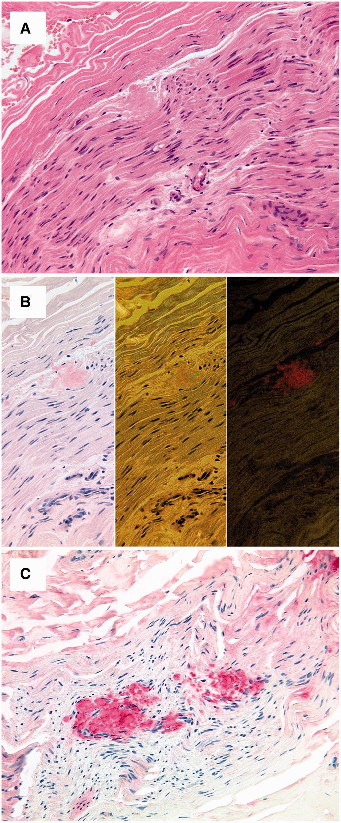
Figure 5.
Histopathology. Sural nerve biopsy of a male patient showing peri- and endoneural amyloid deposits with a homogeneous eosinophilic appearance in haematoxylin and eosin-stained sections (A). Congo red staining yields a pale red staining in bright light (B, left), apple green birefringence in polarized light (B, middle) and an orange fluorescence in fluorescence microscopy (B, right). Immunostaining with an antibody directed against transthyretin shows a strong and even immunoreaction of the amyloid deposits (C).

