
Figure 1.
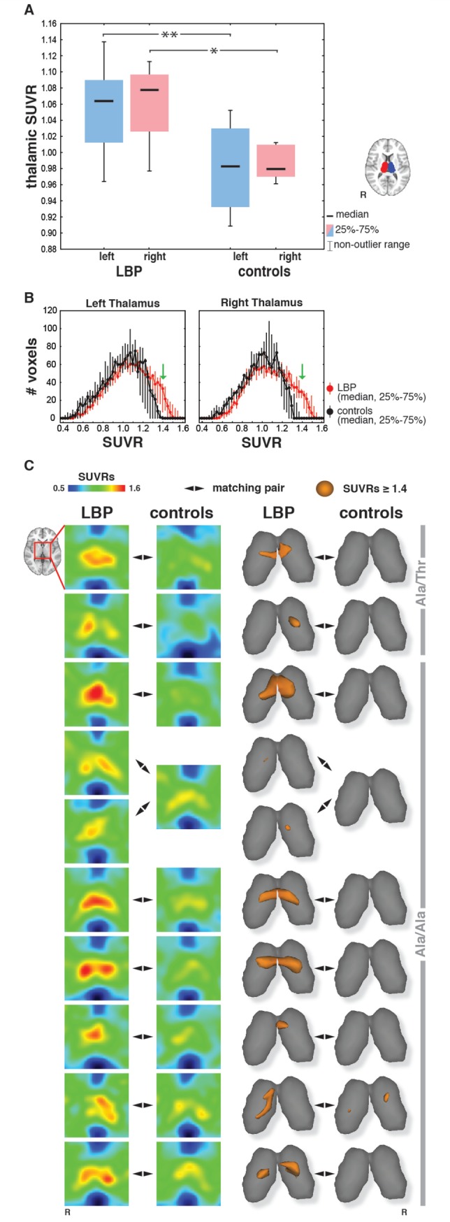
Evidence for glial activation in the thalamus of chronic LBP patients. (A) Boxplots are presented for the mean 11C-PBR28 SUVRs extracted for all 10 patients with chronic LBP and nine control subjects from the thalamic regions of interest (insert). The P-values refer to matched-pairs analyses (sign test) performed using nine chronic LBP-control matching pairs. The analyses were repeated twice, each time using one of the two patients matching the same control, with statistically significant results in both analyses. *P < 0.05, **P < 0.01. (B) Voxel-wise distribution of thalamic SUVRs, showing that patients with chronic LBP have a substantial number of voxels at values ≥ 1.4 (green arrows), whereas controls have a median voxel count of 0. (C) Individual thalamic SUVRs are presented as axial sections (left), and 3D rendering of values higher than the threshold of 1.4 (right). Each row displays SUVRs for each patient-control matched pair. TSPO polymorphism (Ala/Ala or Ala/Thr) is indicated.