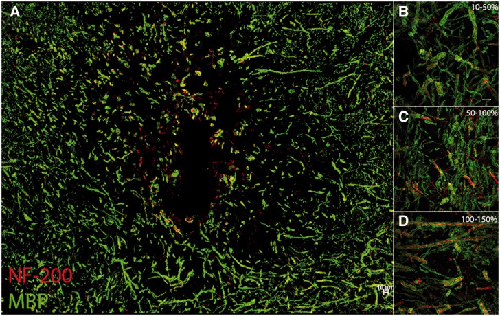
Figure 3.
Molecular disorganization of axons adjacent to lacunar infarcts exists in the absence of significant changes in structural integrity of myelin and axons. Lacunar infarct stained for myelin basic protein (MBP, green) and neurofilament-200 (NF200, red) demonstrate some demyelinated axons immediately adjacent to infarct (A). Note that this image is a serial section of the infarct shown in Supplementary Fig. 1. Higher resolution confocal microscopic images of myelinated axons in increasing distance (B–D) from the infarct show that the structural integrity of axons and myelin remains largely intact despite significant changes in nodal and paranodal length as evidenced by beta-IV spectrin and caspr staining. Tiled confocal image at ×60 (A) and ×100 magnification (B–D). Scale bars = 10 µm.

