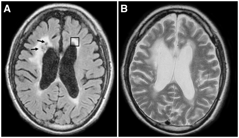
Figure 4.
Representative axial FLAIR and T2-weighted MRI images from one patient with autosomal dominant RVCL. FLAIR sequences demonstrate multiple periventricular lacunar infarcts, predominantly in the right frontal periventricular white matter (arrows, A) along with more diffuse FLAIR hyperintensity within the same regions. T2-weighted images demonstrate both CSF-signal intensity lacunar infarcts as well as hyperintense signal change within the periatrial white matter (B). Immunofluorescently stained sections were taken from brain blocks corresponding to the left periventricular white matter (box) in A.

