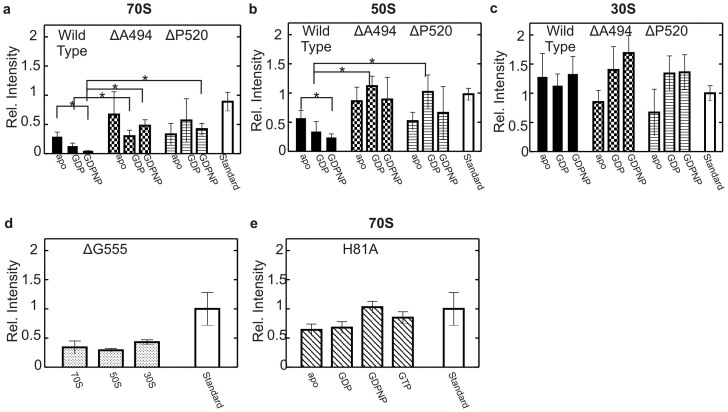
Figure 3. Binding of EF4 and variants to the ribosome in the presence of guanine nucleotides.
The intensity of each band following immunodetection was compared to a 20 pmol standard representing stoichiometric binding. Values with a >85% probability to be significantly different are indicated by a star. 2 μM EF4 wild type (filled), ΔA494 (chequered), ΔP520 (striped) (indicated on top) apo or in the presence of various GDP or GDPNP (0.1 mM) (indicated underneath) bound to (a) 0.1 μM 70S ribosome, (b) 0.1 μM 50S ribosomal subunit, (c) 0.1 μM 30S ribosomal subunits. (d) 10 μM EF4 ΔG555 bound to 0.1 μM 70S, 50S or 30S (indicated underneath) in the presence of GDPNP (0.1 mM). (e) 2 μM EF4 H81A bound to 0.1 μM 70S ribosomes in the presence of various guanine nucleotides (GDPNP, GDP, GTP (0.1 mM)) or apo (indicated underneath).

